
Fig. 4. HPV16 infectivity is increased by knockdown of PIK3C3.
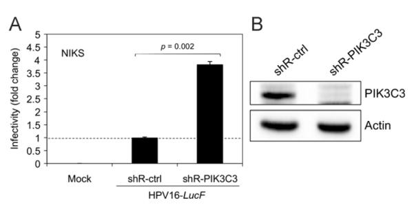
(A) NIKS cells were transduced with lentiviruses containing shRNAs against PIK3C3 (shR-PIK3C3) or a non-target control (shR-ctrl). At 48 h post transduction, the transduced cells were selected by culture in medium containing 1.5 μg/mL of puromycin for 2 weeks to establish stable semi-clonal cell lines. Puromycin-selected cells were inoculated with 10,000 vge/cell of HPV16-LucF and incubated for 48 h. Infectivity was measured and normalized to cell viability as described in Fig. 1. Normalized infectivity data are shown as fold change in infectivity by shR-PIK3C3 over shR-ctrl, and were averaged from quadruplicate samples. Statistical analysis was performed as described in Fig. 3A. (B) In parallel to the infection assay in (A), immunoblotting was performed with the puromycin-selected cells using anti-PIK3C3 (Life Technologies) and anti-Actin antibodies. The data shown here are from one representative of three independent experiments.