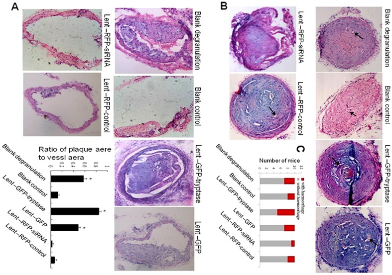
Figure 3. HE stains.
A. HE stain on the uncuffing side of the cervical artery (×100). Compound 48–80 and tryptase increased plaque area and artery stenosis. *P<0.01 vs. blank control, Lent-RFP-siRNA and Lent-RFP-control (n = 5). B. HE stain of the cuffing side of the cervical artery (×100). Tryptase promoted plaque haemorrhage and angiogenesis distinctively. Plaque haemorrhage: red blood cell extravasation directed by black arrows. The cuffing-side artery stenosis was serious, being greater than 90% in all groups, except the lent-RFP-siRNA group. C. Number of mouse with plaque haemorrhage. 50% of mice in Lent-GFP- tryptase group have plaque hemorrhage, while only 1 mouse in siRNA group. *P<0.01vs other groups. (n = 10).

