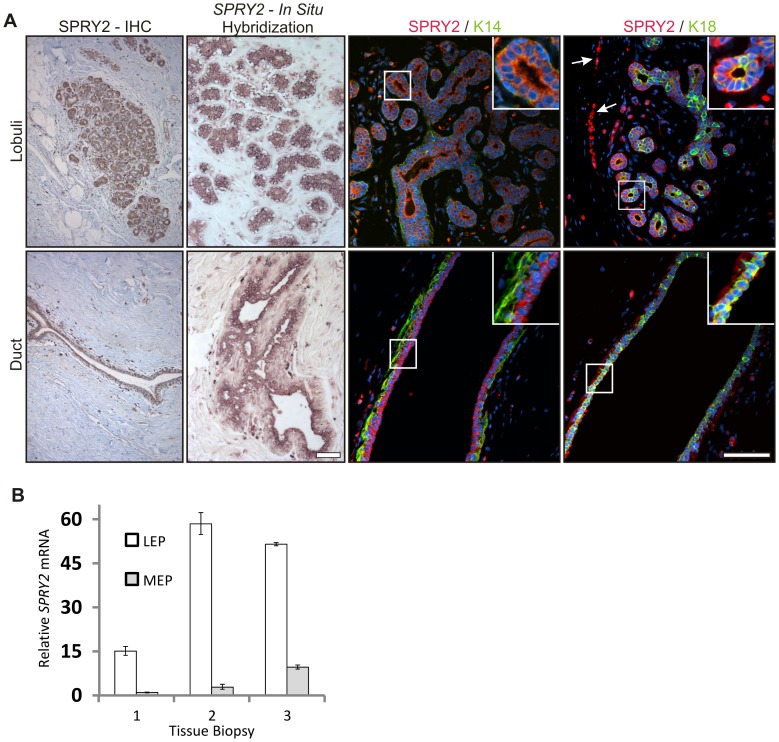
Figure 1. Expression of SPRY2 in lobules and ducts in the normal human breast gland.
Expression of SPRY2 was evaluated in normal human breast tissue derived from reduction mammoplasty biopsies. A) Expression of SPRY2 is most prominent in the luminal epithelial cells. SPRY2 expression was predominantly found within the epithelial compartment of duct and lobuli as evidenced by immunohistochemistry and in situ hybridization. SPRY2 was predominantly expressed in luminal epithelial cells both in ducts and lobuli. SPRY2 was co-stained for K14 (myoepithelial cells) and K18 (luminal epithelial cells). Note the co-expression of SPRY2 and K18 in luminal epithelial cells. SPRY2 expression was also presence in the stroma, most likely in endothelial cells (arrows). Sections were counterstained with TOPRO-3. Bar = 100 µm. B) Expression differences of SPRY2 in luminal- and myoepithelial cells. Real time PCR was used to quantify expression difference of SPRY2 between luminal- and myoepithelial cells. SPRY2 expression was generally low in myoepithelial cells compared to luminal epithelial cells that expressed up to 58 fold more SPRY2. Measurement was done in paired luminal and myoepithelial cells from three different biopsies.

