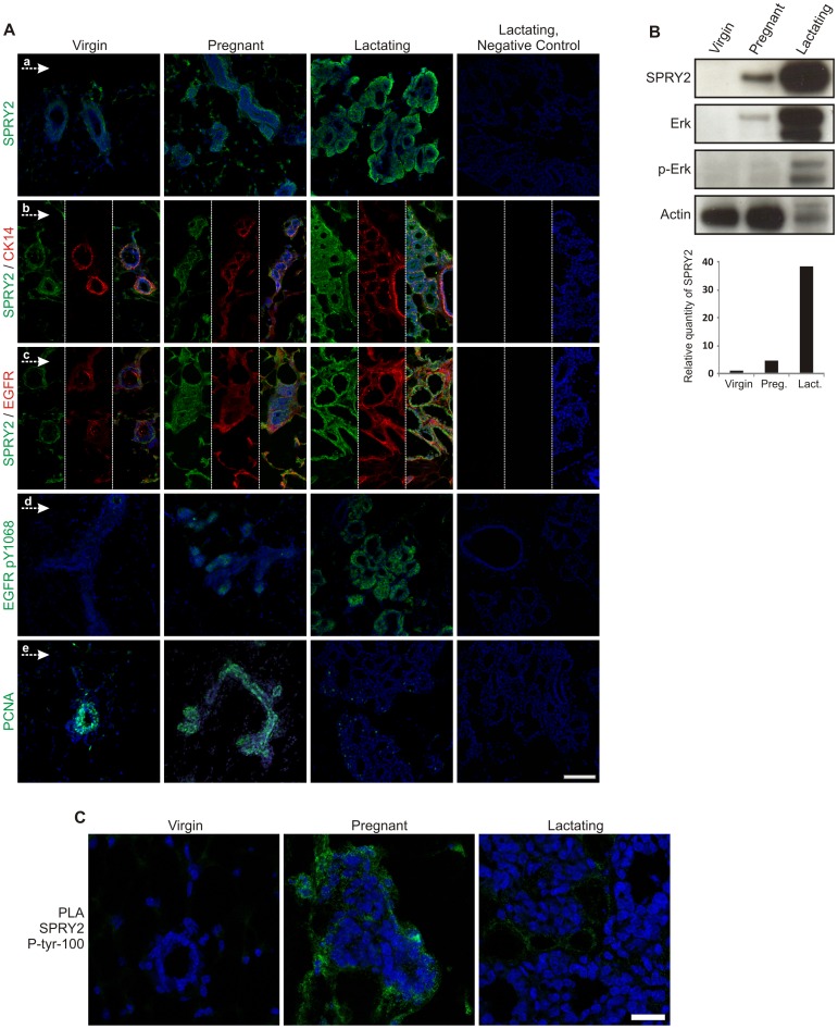
Figure 2. Expression of SPRY2 in virgin, pregnant and lactating mouse mammary gland.
A) Expression of SPRY2 and pEGFR is inversely correlated with cell proliferation. Low expression of SPRY2 is found within the virgin gland with few positive stromal cells (a). Note, increased stromal expression of SPRY2 in pregnant gland accompanied with expression in myoepithelial cell as evidenced by double staining of SPRY2 and the myoepithelial marker CK14 (b). Dramatic increase in SPRY2 expression is seen during lactation (a and b). SPRY2 and EGFR show similar expression pattern at all stages (c) with pEGFR expression seen at terminal buds in pregnant gland. Dramatic increase in pEGFR expression is seen in the lactating gland. Similar expression is found for SPRY2 and pEGFR in lactating gland. Proliferation is increased from virgin to pregnant gland but is reduced during lactation, with only few PCNA positive cells left. Cells counterstained with TOPRO-3, Bar = 100 µm. B) SPRY2 expression is highest during lactation accompanied by activation of Erk/MAPK pathway. Western blot demonstrated the expression differences of SPRY2 in virgin, pregnant and lactating glands. There is over 38 fold increase in SPRY2 expression during lactation compared to virgin state. Total ERK and pERK is also significantly increased during lactation. Actin was used as a loading control. C) SPRY2 activity is peaking during pregnancy in the mouse mammary gland. Using proximity ligation assay it was shown that phosphorylated SPRY2 was significantly more expressed during pregnancy compared to virgin and lactating gland. Bar = 25 µm.

