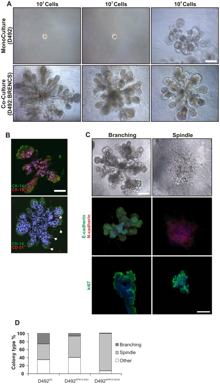
Figure 6. Epithelial integrity is disturbed in SPRY2 KD cells when co-cultured with endothelial cells.
A) Endothelial cells stimulate growth of D492 cells. When plated in 3D rBM culture with breast endothelial cells (BRENCs), D492 cells can form complex branching colonies from as little as 100–1000 cells compared to 7×10³ –104 in 3D monoculture. B) D492-derived branching structures form bi-layered epithelium with BRENCs positioned extralobular. The branching colonies are bi-layered and polarized structures as evidenced by the expression of the myoepithelial marker CK14 on the outer side and the luminal epithelial marker CK19 on the inner side (upper figure). In co-cultures endothelial cells stay as single cells positioned outside the branching structures as seen with CD31 staining (lower figure, arrows). Bar = 100 µm. Sections counterstained with TOPRO-3 nuclear stain. C) Phenotypes of D492 in co-culture with BRENCs. In co-culture with endothelial cells D492 cells form both branching- and spindle-like colonies. The branching colonies show strong expression of E-cadherin, while spindle like colonies have undergone EMT as evidenced by cadherin switch from E- to N-cadherin. Staining for the proliferation marker ki67 shows that both the branching and spindle like colonies are viable and growing. Bar = 100 µm. Sections counterstained with TOPRO-3 nuclear stain. D) Spry2-KD cells show an increase in the spindle-like morphology. While D492NS cells form about 40% spindle-like colonies there is a significant increase in the D492Spry2-KD3 cells up to 65%. The D492Spry2-KD3A form almost exclusively spindle-like colonies in co-culture with endothelial cells.

