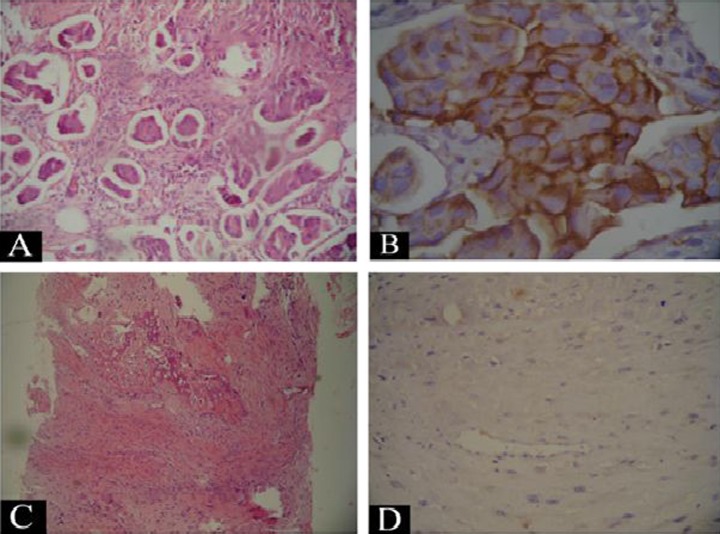
Figure 2.
(A) The tumor area from the breast mass showing invasive groups of malignant moderately differentiated ductal cells (x), (B) the same tumor area showing positive immunostaining of the malignant ductal cells for Her 2 neu (+3) (×200), (C) a microscopic picture of the bony lesion showing malignant spindle cells associated with deposition of osteoid, which supports the diagnosis of osteosarcoma, and (D) negative immunostaining for cytokeratin CK in the malignant osteoblasts.

