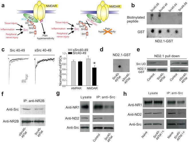
Fig. 1.
Src40-49Tat suppresses the Src-NMDAR interaction in vitro and in vivo. (a) Cartoon illustrating the main hypothesis. Domain structure of Src shows Src-homology (SH) domains 1,2,3 and the unique domain (UD). (b) Dot blot of ND2.1-GST fusion protein probed with biotinylated Src unique domain peptide with overlapping sequence of 40-58, followed by streptavidin-HRP (SA-HRP). (c) Effect of Src 40-49 on mEPSCs. Left panel, average mEPSCs in the first two min after breaking in to whole-cell configuration was superimposed with averages obtained after 8–14 min. Right panel: the ratio of measurements during the first two min and from 8–14 min (Src40-49 n = 11; sSrc40-49 n =5). (d) Dot blot of ND2.1-GST fusion protein probed with biotinylated Src40-49Tat or scrambled Src40-49Tat followed by SA-HRP. (e) In vitro binding assay of assays with ND2.1-GST and Src unique domain, with no peptide, Src40-49Tat or sSrc40-49Tat (30μM). Src unique domain bound to ND2.1-GST was probed with antibody against Src, stripped and reprobed with antibody against GST. (f–h) Immunoblots of coimmunoprecipitates obtained with antibody against NR2B (anti-NR2B, f) or antibody against Src (anti-Src, g–h) from brain crude synaptosomes (f–g) in vitro incubated with Src40-49Tat or sSrc40-49Tat (10μM) or (h) from animals with or without Src40-49Tat (100pmol g−1) intravenous injection (45 min before sample collection). Blots were probed with respective antibodies as labeled.

