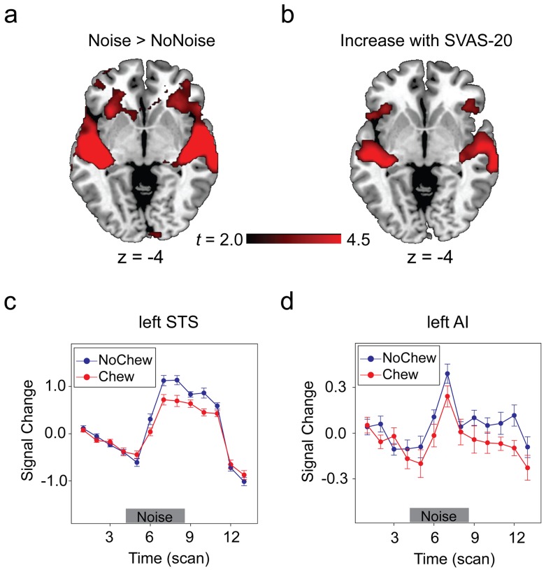
Figure 3. Brain regions revealed by the factorial and parametric models.
(A) Brain regions sensitive to noise and noise-induced stress (“Noise > NoNoise”). (B) Brain regions in which the activation level positively correlates with the ratings of the subjectively experienced level of stress. (C) and (D) The time course of BOLD signal change in the left STS and the left AI reflecting the effect of noise (“Noise > NoNoise”) in the Chew and NoChew conditions. Error bars indicate the standard error of percent signal change (±SEM). To see more clearly the activations in insula, regions illustrated here used a voxel level threshold of p<0.005 (uncorrected) and a extent threshold of 200 contiguous voxels.

