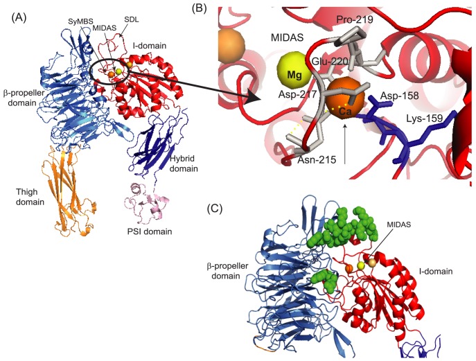
Figure 7. αIIbβ3 headpiece structure and adducted residues.
(A) The X ray crystal structure of αIIbβ3 headpiece was obtained from protein data bank (PDB; 3FCS). There are three metal binding sites in the I domain of the β subunit. Mg2+ in the MIDAS (this site is directly involved in ligands binding) is shown in yellow sphere, while Ca2+ in the SyMBS and ADMIDAS are shown in orange and light orange spheres, respectively. (B) The blowout of residues around metal binding sites from Figure 7 (A) is shown. The adducted residues of photolabeling experiments are shown in blue. Again, Mg2+ in the MIDAS is shown in yellow sphere, while Ca2+ in the SyMBS is shown in orange sphere. Both figures were created using PYMOL. (C) The structure of αIIbβ3 in the open conformation was obtained from Protein data bank (http://www.rcsb.org/pdb/home/home.do; PDB 3FCU). Residues shown as green spheres on αIIbβ3 are suggested PAC-1 binding sites by Puzon-McLaughlin et al. [44]. This figure was created using PYMOL.

