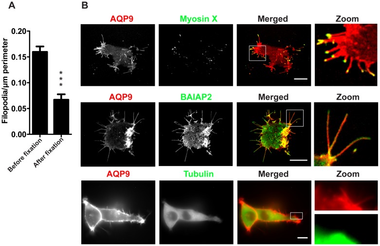
Figure 2. Localization of tubulin, myosin X and BAIAP2 in GFP-AQP9-transfected HEK-293 cells.
(A) Quantification of peripheral filopodia in GFP-transfected HEK-293 cells before and after fixation. Data are presented as mean (± SEM, n = 12–43 cells/group). (B) Images captured at the basal part of a HEK-293 cells co-expressing AQP9 and other filopodia-associated proteins fused to GFP or tagRFP. Linear intensities have been adjusted to visualize the relative distribution of both fluorophores. The zoom panel illustrating AQP9 and tubulin is split to emphasize the lack of tubulin in filopodia. Scalebar 10 µm.

