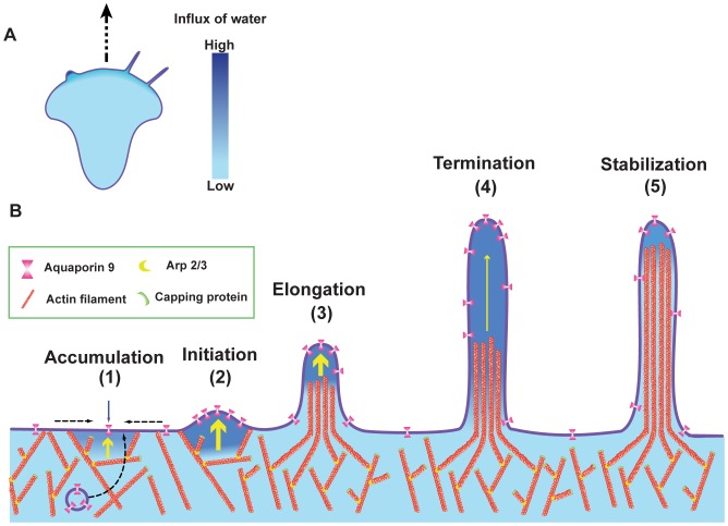
Figure 8. Model for AQP9-induced membrane protrusion.
(A) A migrating cell with lamellipodia, filopodia, and blebs where an increased influx of water corresponds to a darker blue tone. (B1) Local accumulation of AQP9 by vesicle transport and/or lateral membrane diffusion enables a localized increased influx of water across the cell membrane. The influx is driven by an osmotic gradient, likely created by the transmembrane ion distribution (not shown). (B2) The rapid influx of water creates a localized hydrostatic pressure between the membrane and the cytoskeleton pushing the membrane outwards, thus initiating a membrane protrusion. (B3) The influx of water increases the hydrostatic pressure locally. In parallel, actin polymerization is promoted by the exposure of previously membrane-anchored barbed ends and the rapid diffusion of actin monomers in the now diluted, less viscous cytoplasm leading to an elongating filopodium. (B4) Then the rapid water-induced elongation reaches a critical distance from the actin, resulting in termination of the filopodial elongation likely due to equilibration of the water along the filopodium and loss of counter-pressure obtained from the actin cytoskeleton. (B5) The rate of the actin polymerization catches up with the water-induced protrusion and thereby stabilizes the structure. Based on the rate of water flux and equilibration, the filopodium can either protrude once more, or remain at its present length.

