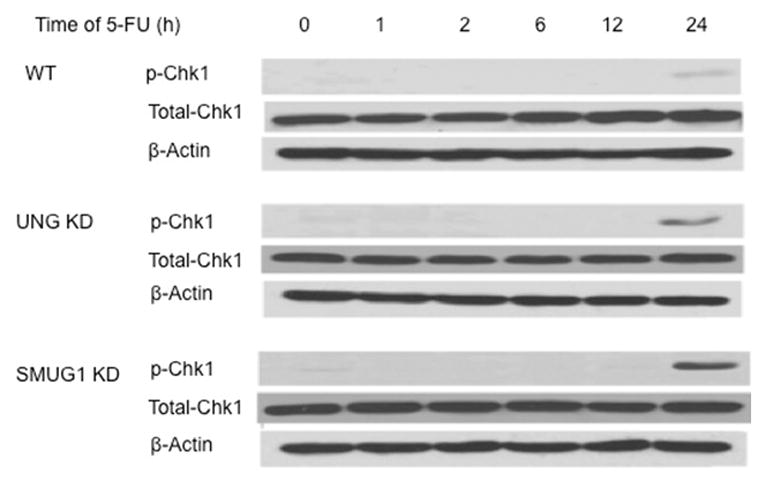
Figure 4.

Western blot analysis of p-Chk1 (Ser345) and total Chk1 protein in whole cell lysates of WT (top panels), LN428/UNG-KD (middle panels), and LN428/SMUG1-KD (bottom panels) cells following treatment with 50 μM 5-FU for the indicated times. β-Actin was used for the loading control. The experiment shown is representative of 3 independent experiments.
