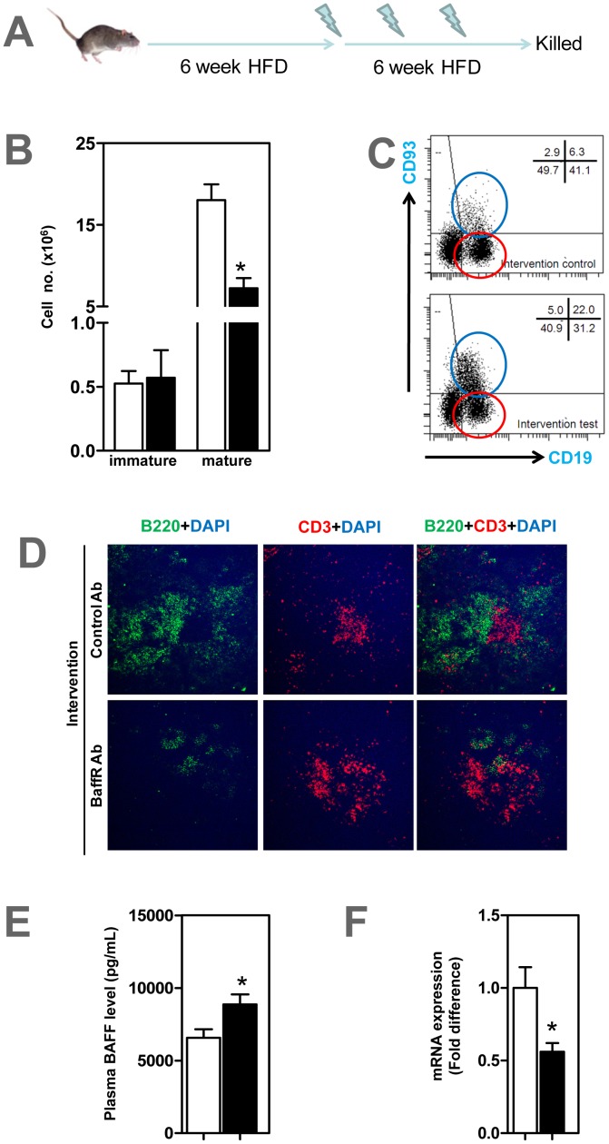
Figure 5. Anti-BAFFR antibody selectively depletes mature B cells, not B1a cells in ApoE−/− mice with established atherosclerosis.
(A) ApoE−/− mice fed a HFD for 6 weeks to generate atherosclerosis, followed by three doses of anti-BAFFR antibody while feeding a HFD for a further 6 weeks. At end of experiment, FACS analysis show (B-C) mature B2 B cells in spleens selectively depleted. Representative FACS image show (C) mature and immature B cells in spleen stained with CD93 and CD19 antibodies Blue circle represents CD93+ CD19+ immature B cells and red circle CD93− CD19+ mature B cells. (D) Spleen B cell zones disrupted in BAFFR-depleted mice compared to control group. (E) Plasma levels of BAFF determined by ELISA increased in anti-BAFFR-antibody treated ApoE−/− mice. (F) mRNA expression of CD20 in spleens reduced in BAFFR-treated mice. n = 7–9 mice per each group * p<0.05 □ control IgG treated mice; ▪ BAFFR-antibody treated mice.

