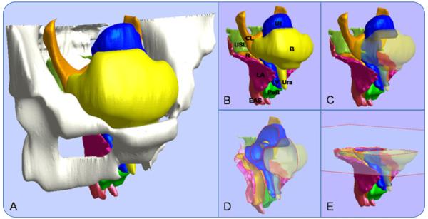
FIGURE 1.
The user can manipulate the 3-dimensional model of pelvic structures. A, A three-quarter right anterolateral view. B, Hiding the bones reveals selected features. C, Making the bladder and urethra transparent reveals the underlying structures. D, Sample sagittal cross-section of the remaining structures. E, Sample axial cross-section.
B, bladder; CL, cardinal ligament; EAS, external anal sphincter; LA, levator ani; PeB, perineal body; R, rectum; Ura, urethra; USL, uterosacral ligament; Ut, uterus; V, vagina.
Luo. A model patient: female pelvic anatomy viewed in diverse 3-dimensional images. Am J Obstet Gynecol 2011.

