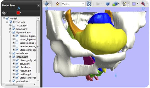
FIGURE 2.
The manipulation interface includes a model tree and toolbar. A, The model tree allows users to hide, isolate, or render transparent individual anatomic structures by right clicking on the label. B, Using this button in the 3-dimensional toolbar at the top, it is possible to zoom in or out, rotate the model, spin it, and pan over it. C, The model tree can be toggled on and off with this button. D, Cross-sections can be cut at a given location and orientation. E, The 3-dimensional model is activated by clicking on the portable document format file.
Luo. A model patient: female pelvic anatomy viewed in diverse 3-dimensional images. Am J Obstet Gynecol 2011.

