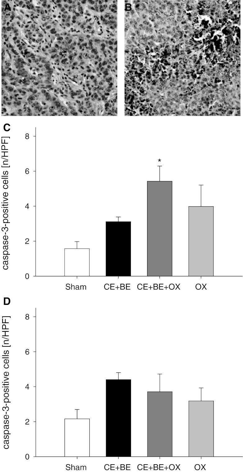
Fig. 3.
Immunohistochemical sections of cleaved caspase-3 as an indicator of apoptotic cell death (a and b). Apoptotic cells are stained brown (a sCHT CE + BE + OX, b HAI CE + BE + OX). Panels c and d display the quantitative analysis of cleaved caspase-3-positive cells in the tumor (given as number per HPF) of animals undergoing HAI (c) or sCHT (d) of cetuximab plus bevacizumab (CE + BE), oxaliplatin (OX) or the combination of all three drugs (CE + BE + OX). Animals undergoing HAI or systemic application of saline served as controls (sham). Data are given as mean ± SEM; *p < 0.05 versus sham. ×200 magnification

