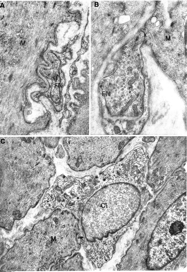
Figure 2.

(A,B) Gastric muscle coat. A nerve ending (FN) is shown in (A) at a distance of about 1000 Å from a muscle cell (M) with a single basal lamina interposed; ×17000. A close neuromuscular junction (asterisk) between a nerve fiber (FN) and a muscle cell (M), with no basal lamina interposed, is shown in (B) ×19,500. (C) Oesophageal muscle coat. Ci: interstitial cell with cytoplasm rich in smooth endoplasmic reticulum. The cell virtually lacks a basal lamina. The arrow points to a close contact with a muscle cell (M). ×8600.
