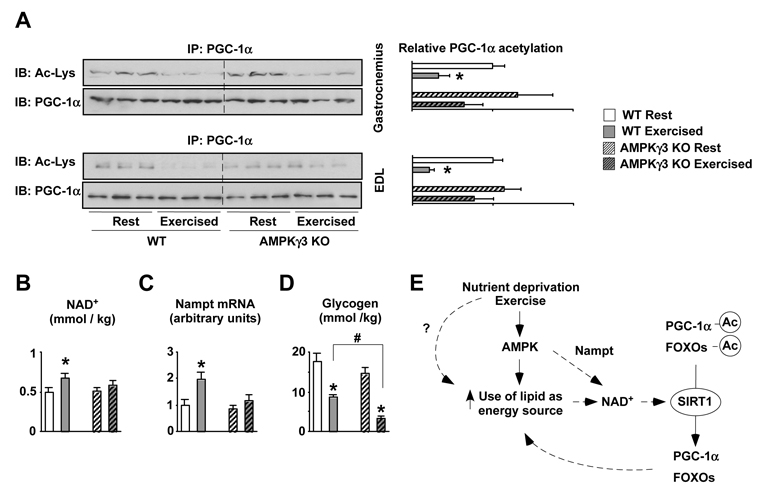
Figure 4. Impaired PGC-1α deacetylation in response to exercise in AMPKγ3 KOs.
(A) Wild-type and AMPKγ3 KO mice were sacrificed at rest or 2.5hrs after swimming (see methods) and muscles were frozen. 300μg of nuclear extracts from gastrocnemius or 2mg of total proteins from EDL muscles were used to immunoprecipitate PGC-1α and check its acetylation levels. (B) Acidic extracts from 50mg of gastrocnemius were used to measure NAD+. Results are shown as mean±S.E. from 3-4 muscles per group measured in duplicate. (C) 20mg of quadriceps were used to measure Nampt expression. (D) 20mg of quadriceps muscle were used to measure glycogen content. Results are shown as mean±S.E. from 3-4 muscles per group measured in duplicate. # indicates statistical difference between the groups indicated (E) Proposed scheme for the coordinated regulation of lipid utilization during energy stress. Upon low nutrient availability or increased energy demand, as during exercise, there is an increase in the AMP/ATP ratio, which activates AMPK, enhancing lipid oxidation in the mitochondria and inducing Nampt levels. These events will raise intracellular NAD+ levels, triggering SIRT1 activation, which deacetylates PGC-1α and FOXOs, both of which regulate genes that further favour mitochondrial respiration and lipid mobilization. Through the figure, * indicates statistical difference vs. respective fed group.

