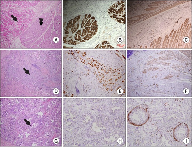
Fig. 4.
Hematoxylin/eosin staining and immunostaining of the anal sphincter. (A-C) Normal anal sphincter. Normal outer striated muscle fibers (arrow) and internal smooth muscle layer (arrowhead) are indicated. (D-F) Three months after anal sphincter injury. Immunostaining showed extensive damage to the muscle fibers with cytoplasmic fibrosis and focal interstitial inflammatory cell infiltration (arrow), and atrophy of the muscle fibers. (G-I) Three months after injection of porous polycaprolactone beads containing autologous myoblasts. There was a marked foreign body reaction characterized by the presence of numerous giant cells and foamy macrophages (arrow), with weak staining for α-smooth muscle actin (A, D, G: hematoxylin-eosin staining; B, E, H: immunostaining of myosin heavy-chain; C, F, I: immunostaining of α-smooth muscle actin; A-G, ×100; H and I, ×200).

