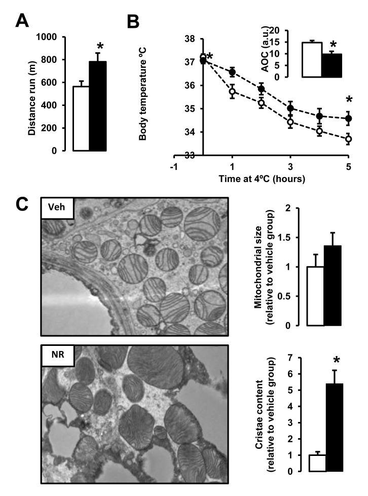
Figure 4. NR enhances skeletal muscle and BAT oxidative function.
10-week-old C57Bl/6J mice were fed a high fat diet (HFD) mixed with either water (as vehicle; white bars and circles) or NR (400 mg/kg/day; black bars and circles) (n=10 mice per group). (A) An endurance exercise test was performed using a treadmill in mice fed with either HFD or HFD-NR for 12 weeks. (B) A cold-test was performed in mice fed with either HFD or HFD-NR for 9 weeks. The area over the curve (AOC) is shown on the top right of the graph. (D) Electron microscopy of the BAT was used to analyze mitochondrial content and morphology. The size and cristae content of mitochondria was quantified as specified in methods. Throughout the figure, all values are shown as mean +/− SD. * indicates statistical significant difference vs. vehicle supplemented group at P< 0.05. This figure is complemented by Fig.S3

