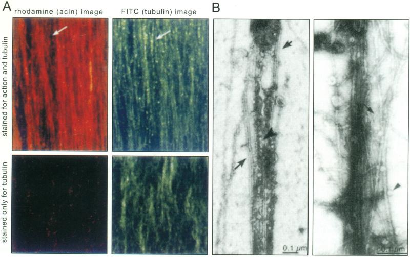
Fig. (1). Actin filaments and microtubules in axoplasm.
A. Actin filaments (red) and microtubules (green) are co-linear in squid axoplasm. Top two panels: extruded axoplasm was fixed and double-labeled with rhodamine phalloidin (left, red) and anti-tubulin (right, green). Note that the two filament systems overlap except in rare areas, such as that indicated by the arrow. Axons stained only for tubulin (bottom panels) do not display any label in the rhodamine channel (bottom left).
B. By electron-microscopy of negatively stained whole mounts of extruded axoplasm, microtubules (large arrows, left panel) and actin filaments (small arrow, right panel) interweave. Membrane-bound vesicles are found attached to either filament type within the plume of interwoven filaments (arrowheads).
(Modified from Bearer and Reese, 1999, J. Neurocytology, by permission).

