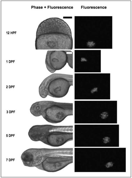
Figure 1.
Persistence of human malignant glioma cells within, and lack of developmental effects on, the developing zebrafish embryo. U251-RFP cells growing within the developing zebrafish embryo for at least 7 d at 30°C do not perturb development. Lateral views of a representative zebrafish embryo after transplantation of U251-RFP human glioma cells, imaged under fluorescence alone (right column images) or merged bright field and fluorescent images (left column images). Leftmost column of text, embryonic age at time of imaging. 12 hpf, embryo imaged at 2 h after transplantation. Bar, 300 μm. The remainder of embryos were imaged at 1, 2, 3, 5, or 7 dpf. Bar, 200 μm.

