
Figure 1.
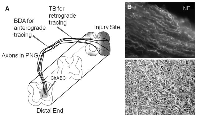
Axon regeneration into peripheral nerve grafts. (A) Diagrammatic representation of a PNG bridging a spinal cord lesion with a demonstration of axonal growth into and through the distal end of the graft where ChABC was delivered to degrade inhibitory matrix molecules. This illustration shows the flexibility of the PNG in that grafts of a specified length can be created, the distal end can be apposed to a specific region or level of the spinal cord and the graft is external to the spinal canal which facilitates isolation for electrophysiological testing and microinjection of tracers into the graft. (B) Regenerating axons immunolabeled with antibody to neurofilament are oriented in a longitudinal array within the PNG. (C) Evaluation of successful grafting includes quantitation of the number of myelinated axons observed in a transverse section through the PNG. Here myelinated axons of various diameters are seen surrounded by fibrous-like connective tissue.