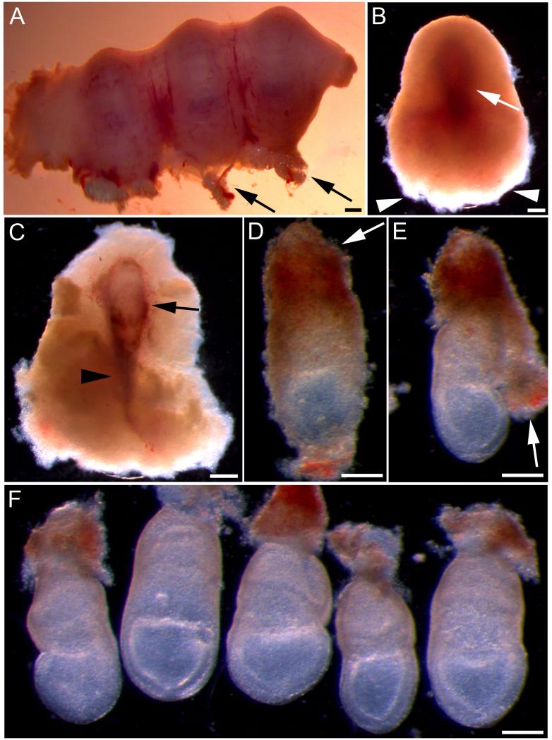
Fig. 1. Dissection of E7.5 mouse embryos for whole embryo culture.
(A) Part of pregnant uterus containing three implantation sites. Arrows indicate fat and blood vessels at the mesometrial surface. (B) Decidual swelling after removal from the uterus. The outline of the embryo can be seen as a darker region (arrow). The next stage of dissection should begin from the broad, fluffy end (arrowheads). (C) Decidual swelling dissected open to reveal the conceptus, encased in trophoblast (arrow). Arrowhead indicates the ectoplacental cone. (D) Conceptus after being released from the decidual swelling. Trophoblast cells can be seen on the surface and the ectoplacental cone remains intact (arrow). (E) Reichert’s membrane and the overlying trophoblast (arrow) have been partially removed by peeling away from the embryonic region. (F) Five dissected embryos after complete removal of Reichert’s membrane. The yolk sacs remain intact and the embryos are now ready to be cultured. Scale bars represent: 400 μm in A-C; 200 μm in D-F.

