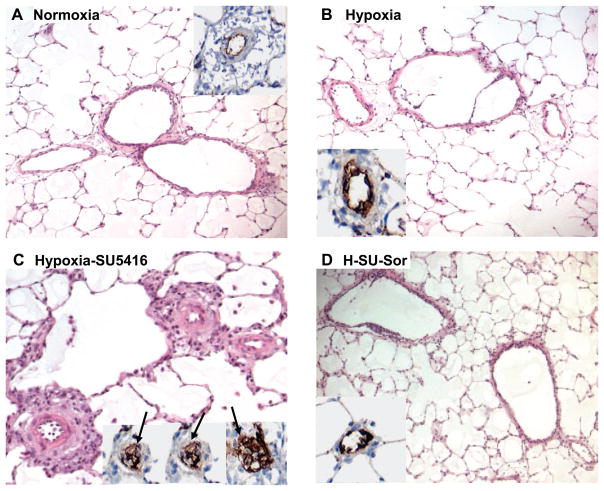
Fig. 3.
Effect of sorafenib on the development of pulmonary hypertension (PH) histopathology. Representative images for each group (hematoxylin and eosin staining) with inset [anti-von Willebrand factor (vWF) staining] demonstrate that compared with normoxic rats (A), rats exposed to hypoxia alone for 3.5 wk displayed only mild lung vascular remodeling (B). In contrast, hypoxia/SU-5416-exposed rats showed marked vascular remodeling with medial wall thickening, endothelial cell hyperproliferation, and formation of plexiform lesions with exuberant vWF-positive endothelial cell proliferation (arrows in 3 representative insets, C). Sorafenib treatment completely prevented the chronic hypoxia-SU-5416-induced vascular remodeling (D).

