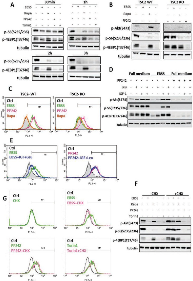
Figure 3.
Activation of lysosomal function is correlated to the suppression of mTORC1. (A) MEFs were treated with EBSS, rapamycin, PP242 or Torin1 (all at 1 μM) for the indicated times. (B) TSC2-WT and TSC2-KO MEFs were treated with EBSS, rapamycin (1 μM) or PP242 (1 μM) for 3 h. (C) TSC2-WT and TSC2-KO MEFs were treated as indicated in panel (B), and cathepsin B enzyme activity was measured as described in Figure 2B. (D) MEFs were incubated in full medium, EBSS, or full medium with PP242 (1 μM) for 2 h, followed by the addition of IGF-1 (200 nM), or Leucine (Leu, 0.2 mg/ml) or IGF1+Leu for another 2 h. (E) MEFs were treated as indicated in panel (D), and cathepsin B enzyme activity was measured as described in Figure 2B. (F) MEFs were treated with EBSS, rapamycin, PP242 or Torin1 (all at 1 μM) in the absence or presence of CHX (10 μM) for 3 h. (G) MEFs were treated as described in panel (F) and cathepsin B enzyme activity was measured as described in Figure 2B. Cell lysate was collected and subject to immunoblotting at the end of above treatment.

