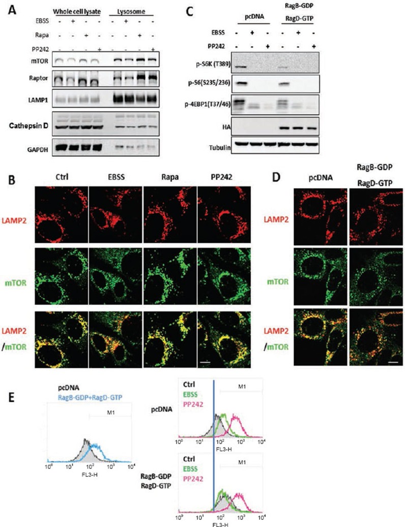
Figure 4.
mTORC1 activity, rather than its lysosomal localization, is critical for its inhibitory effect on lysosomes. (A) MEFs were treated with EBSS, rapamycin (1 μM) or PP242 (1 μM) for 3 h, then lysosome fractions were collected as described in Materials and Methods. LAMP1 was used as a marker for the lysosome fraction. (B) MEFs were treated with EBSS, rapamycin (1 μM) or PP242 (1 μM) for 3 h, and cells were coimmunostained for LAMP2 (red) and mTOR (green). Scale bar, 10 μm. (C) HET293T cells were transfected with pcDNA or the dominant negative RagBGDP-RagDGTP heterodimer for 48 h, then exposed to EBSS or PP242 (1 μM) for 3 h. (D) HET293T cells with the same transfection as described in panel (C), and then coimmunostained for LAMP2 (red) and mTOR (green). Scale bar, 10 μm. (E) HET293T cells with the same transfection and treatment as described in panel (C), and cathepsin B enzyme activity was measured as described in Figure 2B. Cell lysate was collected and subject to immunoblotting at the end of above treatment.

