Figure 7.
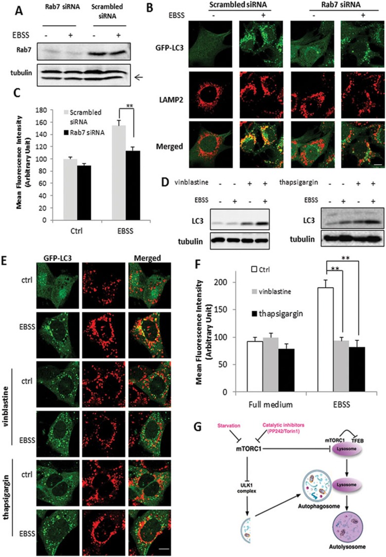
Activation of lysosome function depends on autophagosome-lysosome fusion. (A) MEFs with stable expression of GFP-LC3 were transfected with scrambled or Rab7 siRNA for 48 h. (B) MEFs with or without Rab7 KD were treated with EBSS for 3 h, then processed for LAMP-2 immunostaining (red) to observe the colocalization with GFP-LC3 (green). Scale bar, 10 μm. (C) Following the same treatments as in panel (B), cathepsin B activity was measured as described in Figure 2B. (D) MEFs were pre-treated with vinblastine (20 μM) or thapsigargin (3 μM) for 2 h, followed by EBSS for another 3 h. (E) MEFs with stable expression of GFP-LC3 were subjected to the same treatment as in panel (D) and then processed for LAMP-2 immunostaining (red) and its colocalization with GFP-LC3 (green) was examined. Scale bar, 10 μm. (F) MEFs were treated as indicated in panel (D), and cathepsin B activity was determined as described in Figure 2B. Data are presented as mean ± SD from two independent experiments (**P< 0.01, Student's t-test). (G) Illustration showing the mechanisms mediating the activation of lysosomal function in autophagy involving the mTORC1-TFEB signaling axis and autophagsome-lysosome fusion. Cell lysate was collected and subject to immunoblotting at the end of above treatment.

