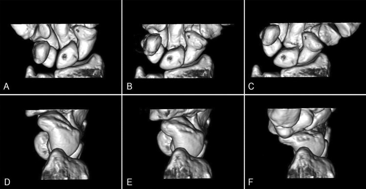
FIG. 3.
Volume-rendered images (top row: palmar view; bottom row: radial view) of a cadaver wrist scanned with a dynamic scanning mode on a dual source CT scanner. Images in ulnar deviation (a, d), neutral (b, e), and radial deviation (c, f) positions show individual carpal bones and joint spaces clearly in three dimensions.

