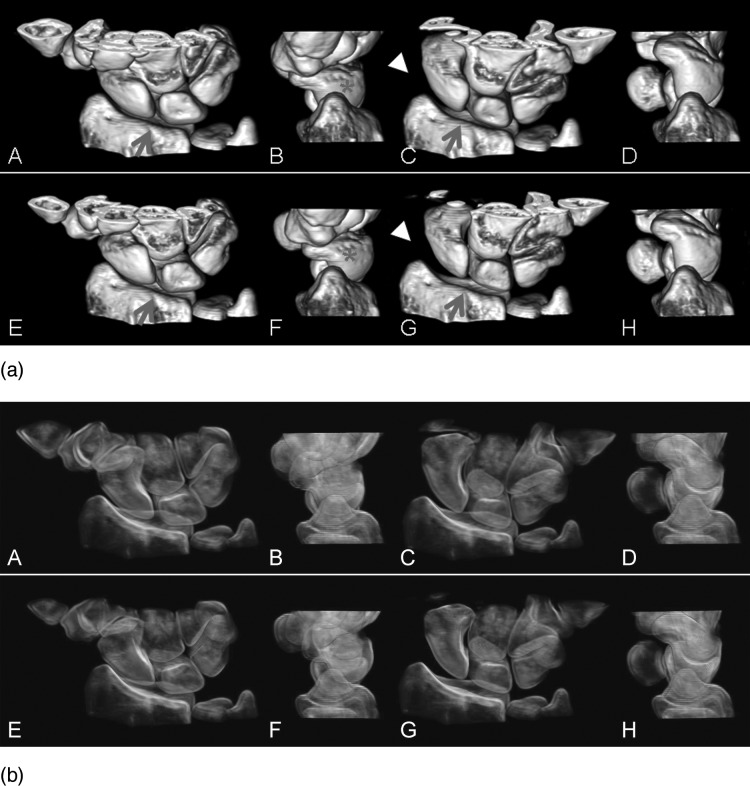
FIG. 5.
(a) Volume-rendered images of a normal wrist (top row) and the wrist after the scapholunate interosseous ligament was cut (bottom row), in radial (A, B and E, F) and ulnar (C, D and G, H) deviation phases during dynamic motion. Both dorsal (A, C and E, G) and radial views (B, D and F, H) demonstrated the deviation, flexion and extension of both the scaphoid and lunate for comparison between the normal and injured wrist. Dissociated motion of the scaphoid and lunate (arrows) can be visualized at both phases in the dorsal views (E and G). Over-flexion of scaphoid (asterisk) during radial deviation in the radial view (F) and increased scapholunate and radioscaphoid distance (arrowheads) during ulnar deviation in dorsal view can be clearly visualized. (b) Volume-rendered images that mimic fluoroscopic images can also be used to better visualize the intercarpal positioning and relative motions.

