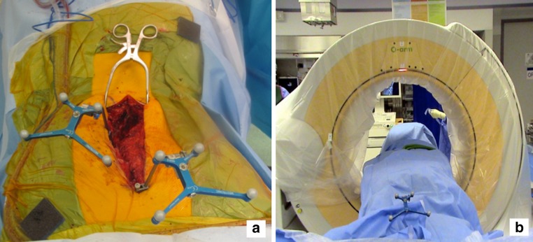
Fig. 1.

a A pair of 3D arrays are attached to the spinous process. The most caudal rays should be placed at or near the apical vertebra, so that the infrared signal from the head of the OR table is not blocked by the most cephalad array. The most cephalad array is placed on the most cephalad spinous process exposed. b After sterile draping the O-arm®, a low radiation CT scan of the intended instrumented vertebrae is performed. Typically, between five and seven vertebrae can be captured successfully on the CT, depending on the size of the patient. Since most spine fusions for scoliosis include between 5 to 14 vertebrae, 2 CT scans are typically required. The information is then fed into the StealthStation®. With experience, the scanning and image preparation typically takes 5–10 minutes
