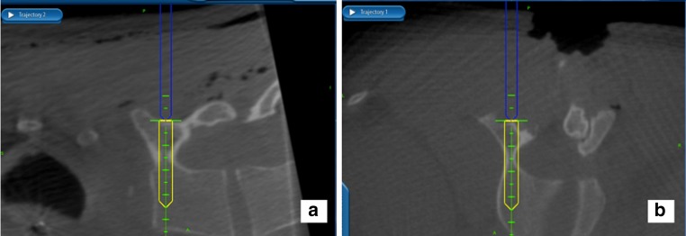
Fig. 3.

The system provides real-time information about the precise length and diameter of the screw that will best fill the pedicle—a crucial feature for instrumenting the variably deformed scoliotic vertebrae. a A sagittal plane image, confirming the optimal cephalad–caudad starting point and sagittal trajectory. b An axial image, confirming the optimal medial–lateral starting point and the largest diameter screw that fits in this pedicle
