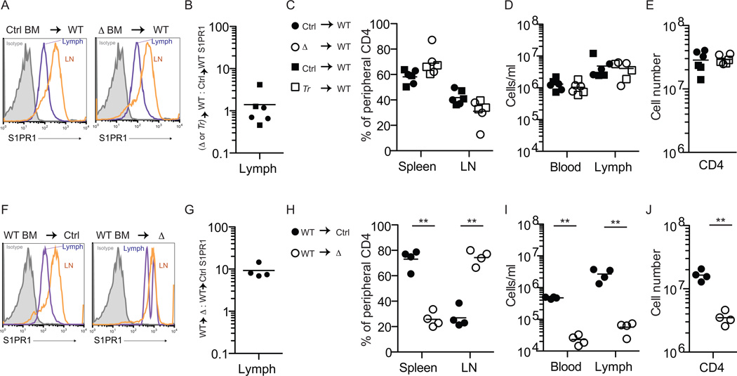
Figure 4. Spns2 is required in endothelial cells to supply lymph S1P and support lymphocyte circulation.
(A-E) Ubiquitin C:GFP+ mice were lethally irradiated and reconstituted with BM from Spns2-deficient mice or littermate controls (GFP+ mice were used to allow assessment of RBC chimerism). Mice were analyzed >6 weeks after transplantation, when RBC and circulating lymphocytes were 97–99% donor-derived. Deletion of Spns2 in Spns2f/fTie2Cre+ (Δ) donor hematopoietic stem and progenitor cells (HSPC) was confirmed by PCR. Data compile 6 pairs of recipients, with 2 pairs of Spns2f/fTie2-Cre+/control BM donors (circles) and 1 pair of Spns2tr/tr (Tr)/control BM donors (squares), analyzed in 6 experiments. (A) Representative histogram of surface S1PR1 on CD62LhiCD4+ T cells in the lymph and lymph nodes of the indicated chimeras. Isotype control is shaded grey. (B) The ratio of surface S1PR1 MFI on CD62LhiCD4+ T cells in the lymph of a WT mouse with Spns2-deficient BM to surface S1PR1 MFI on CD62LhiCD4+ T cells in the lymph of a WT mouse with littermate control BM. (C) Percent of total donor-derived peripheral CD62LhiCD4+ T cells in the spleen and lymph nodes. (D) Total number of donor-derived CD62LhiCD4+ T cells in blood and lymph. (E) Total number of donor-derived CD62LhiCD4+ T cells in the periphery.
(F-J) Spns2f/fTie2-Cre+ mice and littermate controls were lethally irradiated and reconstituted with BM from Ubiquitin C:GFP+ mice. Mice were analyzed >6 weeks after transplantation, when RBC were 88–99% donor-derived and lymphocytes were 85–98% donor-derived. Data compile 4 pairs of mice analyzed in 4 experiments. (F) Representative histogram of surface S1PR1 on CD62LhiCD4+ T cells in the lymph and lymph nodes of the indicated chimeras. Isotype control is shaded grey. (G) The ratio of surface S1PR1 MFI on CD62LhiCD4+ T cells in the lymph of a Spns2f/fTie2-Cre+ mouse with WT BM to surface S1PR1 MFI on CD62LhiCD4+ T cells in the lymph of its littermate control with WT BM. (H) Percent of total donor-derived peripheral CD62LhiCD4+ T cells in the spleen and lymph nodes. (I) Total number of donor-derived CD62LhiCD4+ T cells in blood and lymph. (J) Total number of donor-derived CD62LhiCD4+ T cells in the periphery. *p<0.05, **p<0.01. See also Fig. S3 and Fig. S4.

