Figure 1.

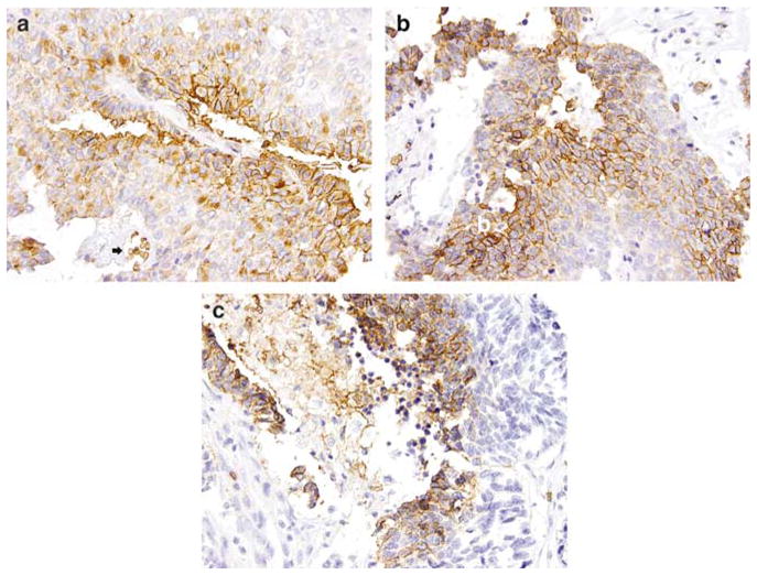
GLUT-1 expression in pulmonary neuroendocrine carcinomas. On immunohistochemical studies, GLUT-1 shows a distinctive membranous staining pattern (a, atypical carcinoid, ×400); the bottom of the field (black arrow) shows a vessel with erythrocytes serving as an internal positive control. The staining is more prominent at the luminal borders of the tumor islands (b, large cell neuroendocrine carcinoma, ×400) or in the areas adjacent to necrosis (c, small cell carcinoma, ×400). GLUT-1, glucose transporter-1.
