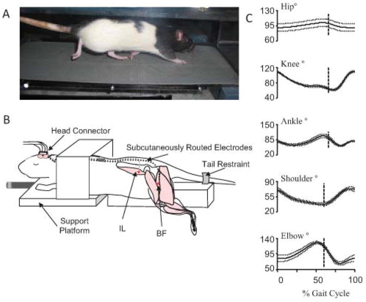
Figure 1.
Rat locomotor testing and therapy. (A) Rat walking on a treadmill, reflective markers are placed on bony prominences for 3D visualization to collect kinematic data. (B) Stimulation therapy setup (figure modified from64). Electrodes for neuromuscular stimulation are implanted in the hip flexors (IL-iliacus) and extensors (BFh- biceps remoralis anterior head). Electrode leads are routed to a head connector for connection to a stimulator. The rat is mounted on a platform for stimulation training. (C) Sham injured kinematic data from a rat walking on a treadmill (data from the study described in71). Note the biphasic nature of the knee and ankle trajectories.

