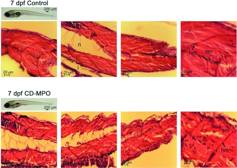
Figure 2. Histochemistry of the musculature in 7 dpf control and CD–MPO zebrafish.
Control and CD–MPO-injected zebrafish at 7 dpf showing an apparent ‘quasi-normal’ phenotype were subjected to longitudinal sectioning and H&E staining of the musculature. A selection of images from three independent experiments is shown. Symbols: hm, horizontal myoseptum; i, intestine; n, notochord; sm, somitic muscle.*syncytial nucleus; **eccentric nucleus.

