Figure 1.
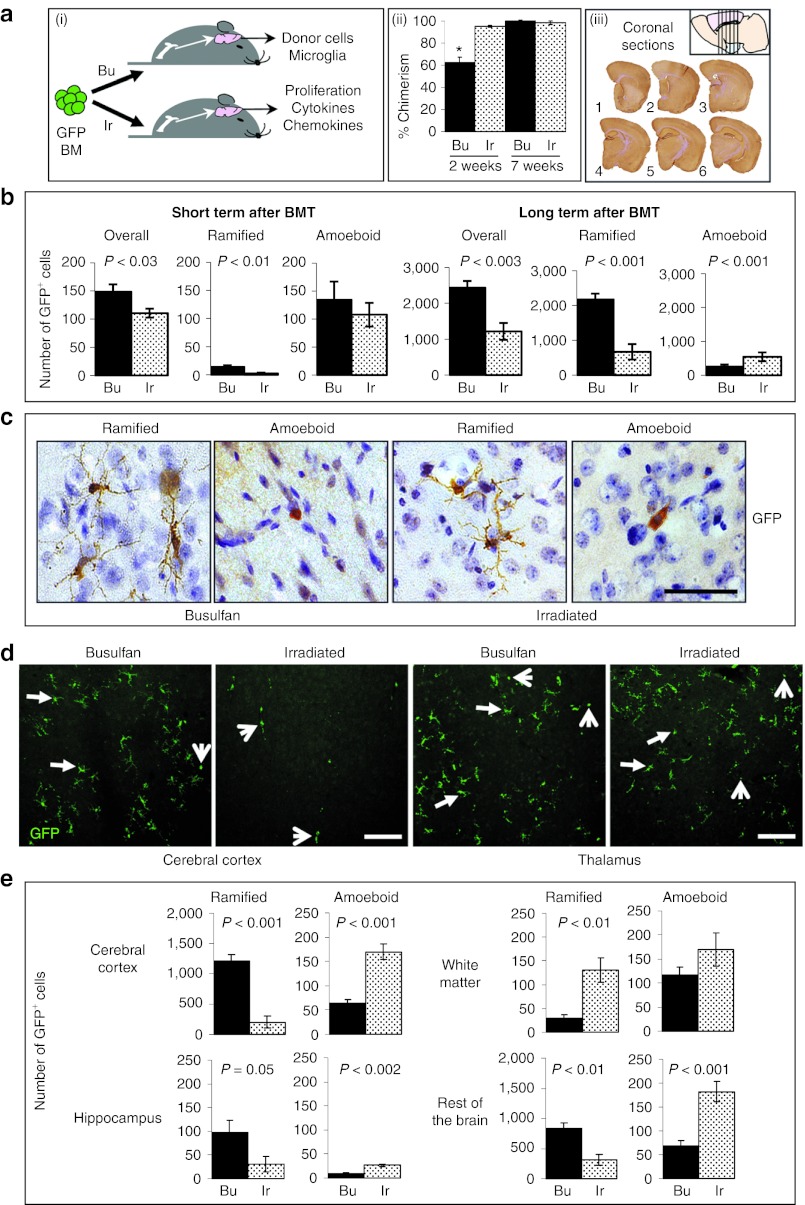
Quantification of total, resting, and activated donor-derived microglia in the brains of busulfan- and irradiation-conditioned transplant recipients. (a) (i) GFP+ bone marrow (BM) was delivered via the tail vein into mice that had undergone full myeloablative conditioning, using either busulfan (Bu) or irradiation (Ir), to investigate the level of brain engraftment after BMT. (ii) Peripheral blood donor cell chimerism (GFP) was significantly lower in busulfan-conditioned recipients compared with the irradiated (*P < 0.0001) 2 weeks after BMT with full chimerism reached in both groups by 7 weeks (n = 6/group). (iii) 6 to 9 months after BMT, the number of GFP+ cells were counted from six coronal sections of brain taken from Bregma 0.98, 0.26, −0.46, −1.18, −1.94, and −2.62 mm (1 to 6; n = 6 mice/group). (b) GFP+ cells (brown; nuclei stained blue) were counted according to their morphology; (c) ramified or amoeboid (bar = 50 µm) in both the short (2 weeks) and long term (6–9 months) after BMT. The overall number of GFP+ cells and number of ramified donor-derived GFP+ cells in busulfan-conditioned brain was significantly higher than irradiated brain at both timepoints (P < 0.03). Irradiated brain contained significantly more activated amoeboid GFP+ donor-derived cells long term after BMT (P < 0.001). (d) Distribution of GFP+ cells (green) in the primary motor, somatosensory and parietal areas of the cerebral cortex and the submedial and ventromedial thalamic nucleus of irradiated and busulfan-conditioned brains taken from the same area of Bregma (−0.46 mm) were visualized using confocal microscopy (bar = 100 µm). Arrows show ramified cells and arrow heads show amoeboid GFP+ cells. Ramified GFP+ cells were more likely to be detected in the submedial and ventromedial thalamic nucleus. (e) Significantly more ramified GFP+ cells were detected in all regions of busulfan-conditioned brain (cerebral cortex, hippocampus, and the rest of the brain; P = 0.05) except in the white matter, long term after BMT. By contrast, significantly more amoeboid GFP+ cells were detected in all regions of irradiated brain (P < 0.001) except in the white matter. Error bars represent the SEM and P values are from Student's t-test (b and e) or one-way analysis of variance with Tukey's multicomparison test (a (ii)). BMT, bone marrow transplantation; GFP, green fluorescent protein.

