Figure 1.
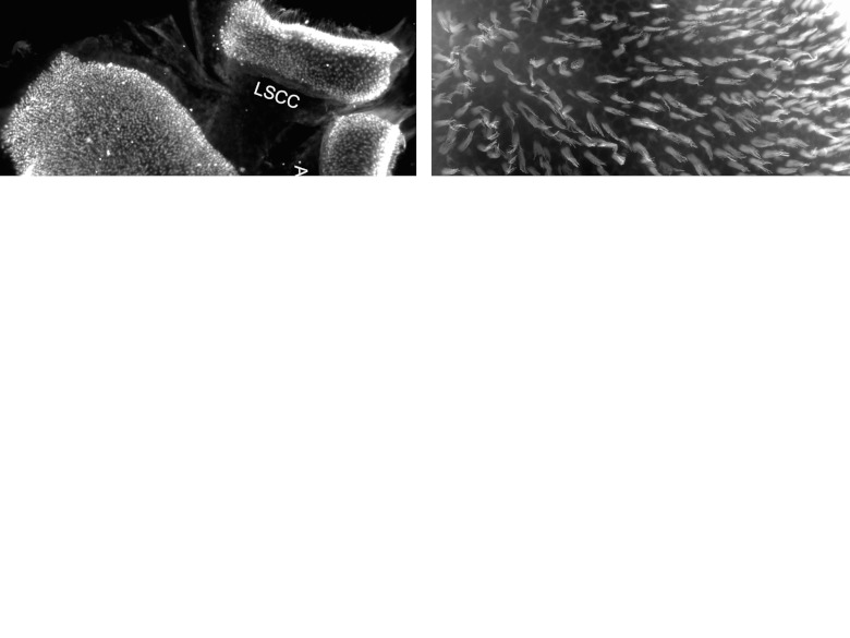
Epi-fluorescence images of vestibular epithelia taken from normal ears (a,b) or an ear obtained 1 week after ototoxic trauma (c,d). (a) The sensory epithelium (SE) of the utricle (on left) and two semicircular canals (LSCC for lateral and ASCC for anterior) on the right, stained for Myosin VIIa, showing dense and even distribution of positive cells, with staining visible in stereocilia bundles. (b) Phalloidin staining in the utricle reveals long stereocilia bundles throughout the sensory epithelium. (c) After the lesion, the number of cells stained by Myosin VIIa appears greatly diminished and the few stained cells exhibit clumps or dysmorphic aggregations in both utricle and canal organs. (d) The contour of adherens junctions stained with phalloidin shows normal appearance in the transitional epithelium (TE) but the sensory epithelium regions lacks hair cell bundles and the apical junctional organization is perturbed. Scale bar: a and c are 50 µm; b and d are 20 µm.

