Figure 3.
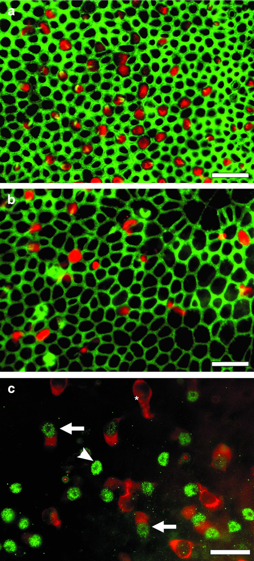
Whole mounts of utricles viewed in epi-fluorescence after ototoxic lesion followed by treatment with Hes5 siRNA (a,c) or control reagent (b). (a) Three weeks following the lesion (2 weeks after Hes5 siRNA injection), anti-Myosin VIIa antibody (red) and phalloidin (green) reveal numerous Myosin VIIa-positive cells in the tissue. These presumed regenerated hair cells appear in variable size and apical contour shape ranging from round to nearly triangular. The stereocilia bundles are viewed in green and appear to have irregular orientation and planar polarity. The density of cells is lower than in normal controls (Figure 1b). (b) Control-treatment sample at same time-point shows few poorly organized Myosin VIIA-positive cells. The density of cells appears lower in the control tissue and the width of the junction actin belt thinner than in the siRNA-treated utricle (a). The diameter of hair cells appears similar in a and b. (c) One week after Hes5 siRNA injection (2 weeks after the lesion) the sensory epithelium region of the utricle, stained for Atoh1 (green) and Myosin VIIa (red) includes Atoh1-positive/Myosin VIIa-negative cells (arrow head), Atoh1-negative/Myosin VIIa-positive cells (asterisk), and cells positive for both (arrow). Scale bar: 20 µm.

