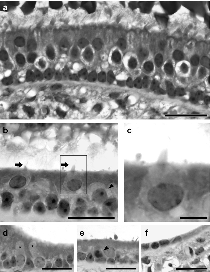
Figure 4.
Light microscope images of cross-sectioned utricles from a normal (untreated) ear (a), or ears after ototoxic lesion followed by treatment with Hes5 siRNA (b–e) or vehicle control (f). (a) The normal utricular sensory epithelium contains hair cells and nonsensory cells. Type I hair cells are surrounded by a calyx. Stereocilia are tall and appear embedded in the otoconial membrane. Supporting cell nuclei are arranged in a orderly line just above the basement membrane. (b) In lesion and then siRNA-treated utricle, hair cells are close to the luminal surface of the epithelium and exhibit short stereocilia bundles (arrow). There were some nuclei in intermediate positions (arrow head). These cells have larger and lighter nuclei than cells in the supporting cell layer. Nuclei in the supporting cell layer are not as well organized near the basement membrane as in the normal utricle. (c) Zoom-in image of highlighted area in b shows one longest cilium. (d) Some nuclei located at the supporting cell level have distinguishing features based on size and density of chromatin, suggesting they are newly transdifferentiating cells (asterisks). (e) Cells containing a darker nucleus are possibly undergoing a degenerative process (arrow head). (f) Cross-section of a utricle observed at same time-point, 2 weeks after vehicle (control) injection. The tissue is devoid of hair cells. All scale bars: 20 µm.

