
Figure 2. Mutational Analysis of TGFB2 in aneurysm patients.
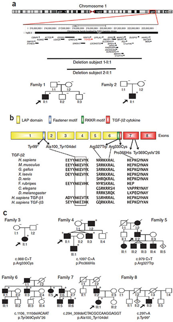
(A.) Schematic representation of the microdeletions on chromosome 1q41. The TGFB2 gene is indicated in red. Pedigrees for two patients with de novo chromosomal microdeleletions completely overlapping TGFB2 (1-II:1 and 2-II:1) are shown. (+) Indicates presence of the described mutation in an associated individual while (−) indicates lack of mutation. (B-C.) TGFB2 mutations and pedigrees for families 3–8. Pedigrees document an autosomal dominant pattern of inheritance. Mutations are annotated at the nucleotide (c.) and protein level (p.; three letter code for amino acids is used; reference transcript: Ensembl ENST00000366929 or NCBI NM_001135599.2). Circle, female; square, male; open symbol, unaffected; shaded symbol, affected; diagonal line, deceased. The location of mutations in relation to the exons (numbers) of TGFB2 and the domain organization is shown (LAP; latency associated peptide; RKKRA potential furin cleavage site). Evolutionary conservation of the mutated residues in TGFB2 and related human cytokines (TGFB1/3) is shown.