
Figure 3. Cardiovascular Pathology in Human Subjects with TGFB2 mutations.
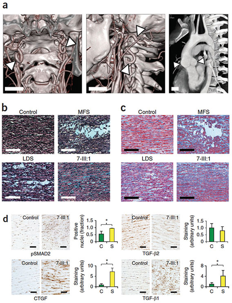
(A.) Multidetector computed tomography (MDCT) with 3 dimensional reconstruction of head and neck vessels demonstrating tortuosity of the distal cervical internal carotid arteries bilaterally (arrows, center panel) as well as the V1 segment of the left vertebral artery (arrows, left panel). MDCT imaging in modified sagittal view of dilated aorta at sinuses of Valsalva (arrows, right panel), Bars= 2 cm. (B.) Movat’s pentachrome staining of human aortic samples demonstrating an increase in proteoglycan deposition (Blue staining in Movat’s pentachrome) and elastic fragmentation (Black in Movat’s pentachrome) in MFS, LDS, and patient with TGFB2 mutation (7:III-1) versus control, Bar= 200µM, Enlargement Bar= 80µM. (C.) Masson’s Trichrome staining of human aortic samples with increased collagen deposition (Blue in Masson’s Trichrome) in MFS, LDS, and patient with TGFB2 mutation (7:III-1) versus control, Bar= 200µM, Enlargement Bar= 80µM. (D.) Immunocytochemical staining of the aortic media for phosphorylated Smad2 protein, CTGF, TGFB1, and TGFB2. Panels show control aorta (Control) and patient aorta with TGFB2 mutation (7:III-1). Quantification of fraction of pSmad2 positive nuclei (pSMAD2) or staining (CTGF, TGFB1, TGFB2) represents staining of three control aortas (Co) versus patients 7:III-1 and 5:II-2 (Pts), Error bars equal 2 SEM, (*p<0.05). Bar= 80µM.