
Figure 5. Synergistic pathology in Tgfb2+/−:Fbn1+/C1039G double heterozygous mice.
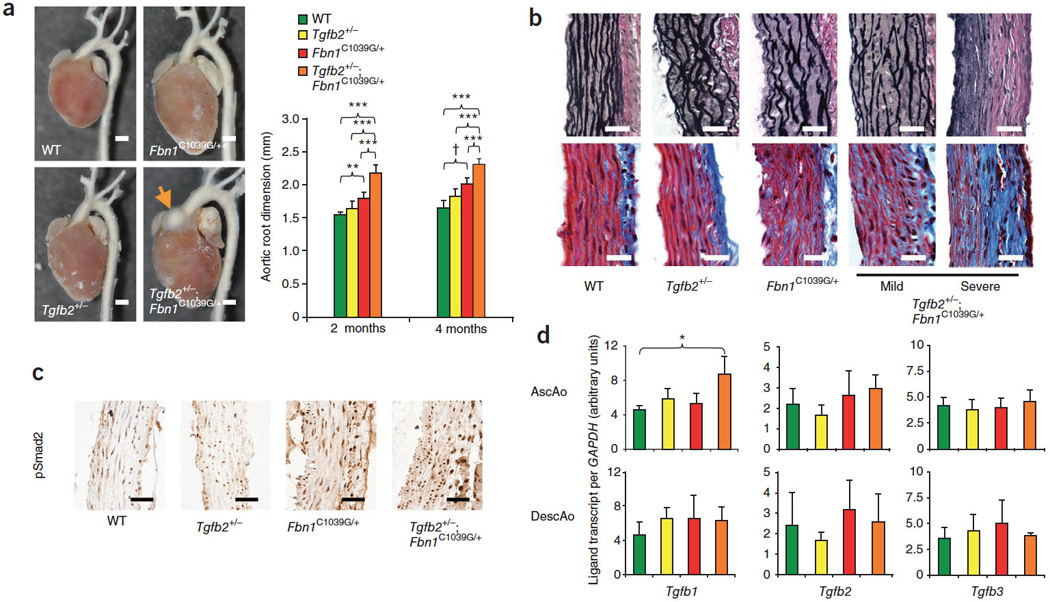
(A.) Photomicrographs and echocardiographic aortic root quantification of WT (n=8), Tgfb2+/− (n=8), Fbn1+/C1039G (n=7), and Tgfb2+/−: Fbn1+/C1039G (n=11) mice. Orange arrow demonstrates a large sinus of Valsalva aneurysm. (**p<0.01, †p<0.005, ††p<0.001) Bar = 1.5 mm. (B.) Worsened aortic phenotype from Tgfb2+/−: Fbn1+/C1039G double heterozygous mice. Panels of VVG (upper row) and Masson’s Trichrome (bottom row) stained aortas from 4 month old mice demonstrating elastin fragmentation and increased collagen deposition in Tgfb2+/−: Fbn1+/C1039G mice. Bar= 20 µm. (C.) Immunohistochemistry of phosphorylated Smad2 in aortas from 4 month old WT, Tgfb2+/−, Fbn1+/C1039G, and Tgfb2+/−: Fbn1+/C1039G mice. Bar= 20 µm (D.) Transcript analysis of ascending and descending aortas of two month old WT (n=3), Tgfb2+/− (n=3), Fbn1+/C1039G (n=3), and Tgfb2+/−: Fbn1+/C1039G (n=3) mice normalized to GAPDH expression, Error bars equal 2 SEM, (*p<0.05).