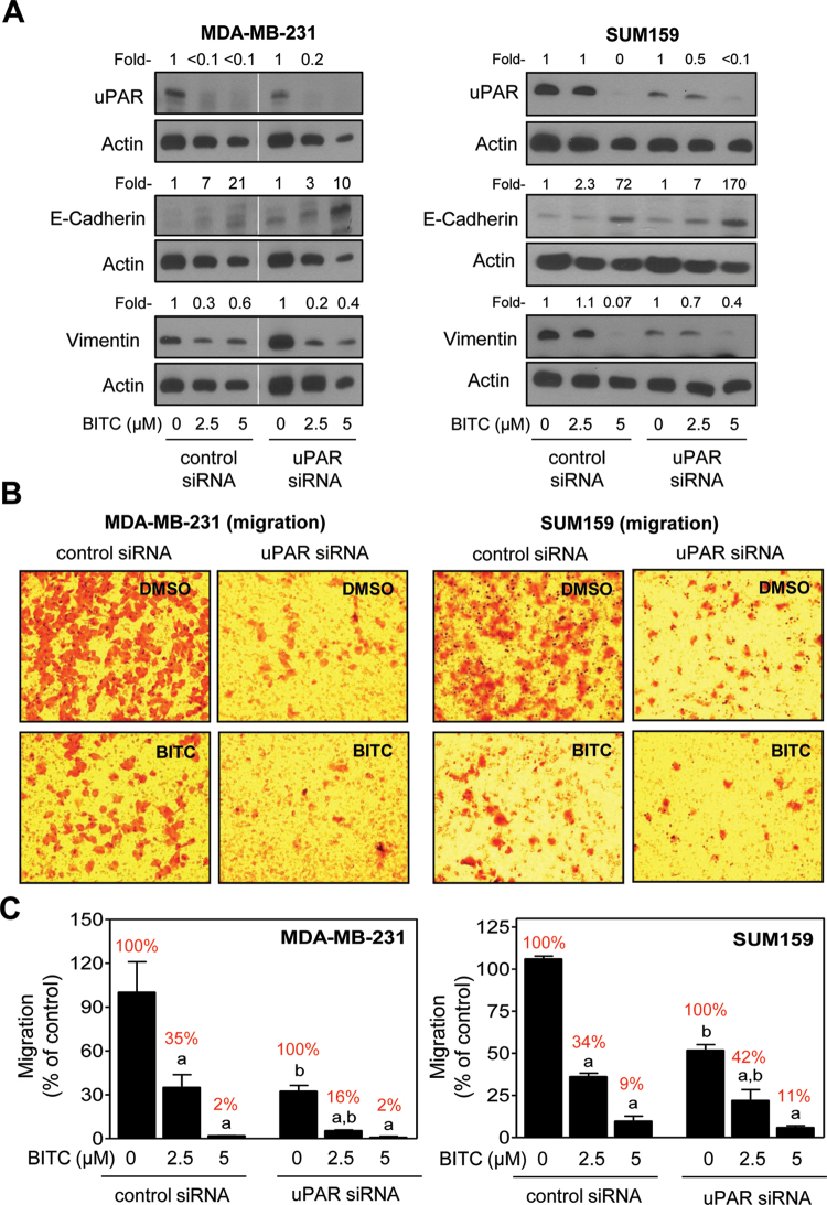
Fig. 4.
Effect of uPAR protein knockdown on BITC-mediated inhibition of EMT and cell migration in MDA-MB-231 and SUM159 cells. (A) Western blotting for uPAR, E-cadherin and vimentin proteins using lysates from MDA-MB-231 and SUM159 cells transiently transfected with a non-specific (control) siRNA or uPAR-targeted siRNA and treated for 24h with DMSO or BITC (2.5 and 5 μM). Numbers above the bands represent densitometric quantitation relative to corresponding DMSO-treated control. (B) Representative microscopic images depicting migration by MDA-MB-231 and SUM159 cells transiently transfected with a non-specific (control) siRNA or uPAR-targeted siRNA and treated for 24h with DMSO or 2.5 μM BITC (×100 magnifications). (C) Quantitation of cell migration from data shown in panel B. Results shown are relative to control siRNA transfected cells treated with DMSO (mean ± SD, n = 3). Percent values in red font represent normalization to corresponding DMSO-treated control for each cell type. Significantly different (P < 0.05) compared with acorresponding DMSO-treated control and bbetween control siRNA and uPAR siRNA transfected cells at each concentration of BITC (0, 2.5 and 5 μM) by one-way ANOVA followed by Bonferroni’s multiple comparison test. Similar results were observed in replicate experiments in each cell line.

