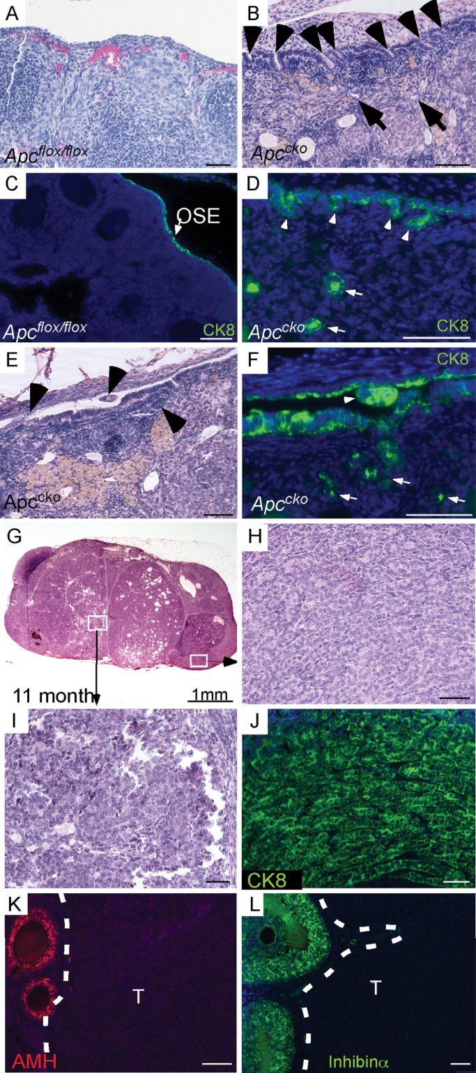
Fig. 1.
Histology of Apccko ovaries. (A) Normal histology of the OSE in older (>4 month) control ovaries by hematoxylin and eosin. (B) Invasion of stromal cells by hyperplastic OSE cells (arrowheads) in Apccko ovaries at 7–9 months. Epithelial gland-like structures (arrows) were observed in ovarian stroma. (C and D) CK8-specific staining showed normal OSE cells in control ovaries (arrow), and hyperplastic OSE cells (arrowheads) and glandular (arrows) epithelial cells in stroma of mutant ovaries at 7–9 months. (E and F) CK8-positive epithelial cells were observed in the space between the OSE and the ovarian bursa (arrowhead). Mutant OSE cells forming epithelial glandular structures (arrows) in mutant ovaries at 7–9 months. (G–I) OEAs formed in mutant ovaries. IF of the Apccko tumors with CK8 (J), anti-Müllerian hormone (K) and inhibinα (L). T = tumor area in K and L at 7–9 months. In panels C, D, F, J, K and L, nuclei are shown stained with 4′,6-diamidino-2-phenylindole (DAPI). Bars: 50 µm.

