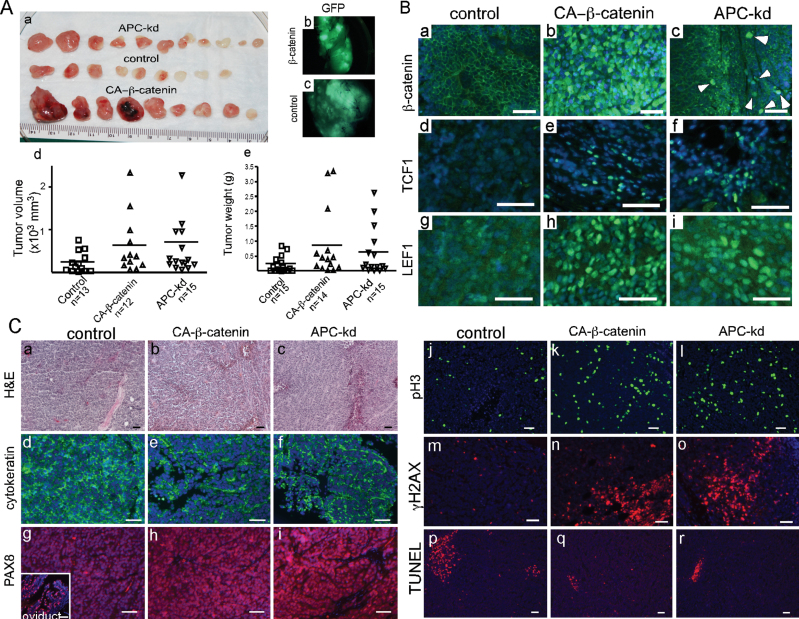
Fig. 4.
Xenotransplants of HOSE cells with either CA-β-catenin or APC knockdown. HOSE cells were transduced with control virus or, virus to express CA-β-catenin or to knockdown (kd) expression of APC. Gross tumors harvested 4 weeks after subcutaneous injection of the transduced HOSE cells into the dorsal flanks of nude mice (A, a). Direct GFP fluorescence in control and CA-β-catenin tumors (A, b and c). Tumor volume and weight in three different groups of tumors (A, d and e). Expression of β-catenin, TCF1 and LEF1 was detected by IF in tumors as indicated (B). Histological and IF (cytokeratin, PAX8, pH3, γ-H2AX and terminal deoxynucleotidyl transferase-mediated dUTP nick end labeling) examination of tumors (C). Inset in panel C, g, is a positive control for PAX8 staining in mouse oviduct. DAPI staining was used to indicate nuclei. Bars: 50 µm.

