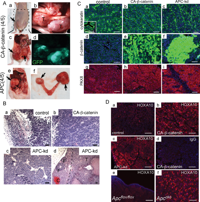
Fig. 5.
Tumor development with intraperitoneal injection of transduced HOSE cells. (A) Gross analysis of tumors formed after intraperitoneal injections of CA-β-catenin (a–d) and APC knockdown (e–f) HOSE cells. CA-β-catenin tumors were GFP positive (d). Histology of control (B, a), CA-β-catenin (B, b) and APC knockdown (B, c and d) tumors. In panel B, d the black dotted line demarcates tumor area of cells that targeted the normal ovary of nude mice; follicles (F). Localization of cytokeratin, β-catenin and PAX8 in tumors from all three groups of animals by IF (C). Mouse oviduct was used as positive control for cytokeratin localization (inset in panel C, a). HOXA10 protein expression in control (D, a), CA-β-catenin (D, b) and APC knockdown (D, c) HOSE tumors. (D, e and f) HOXA10 expression in Apccko ovarian tumors and controls. (D, d) Negative control for HOXA10-specific staining with an equal amount of non-immune IgG. Nuclei are stained with DAPI. Bars: 50 µm.

