
Figure 4.
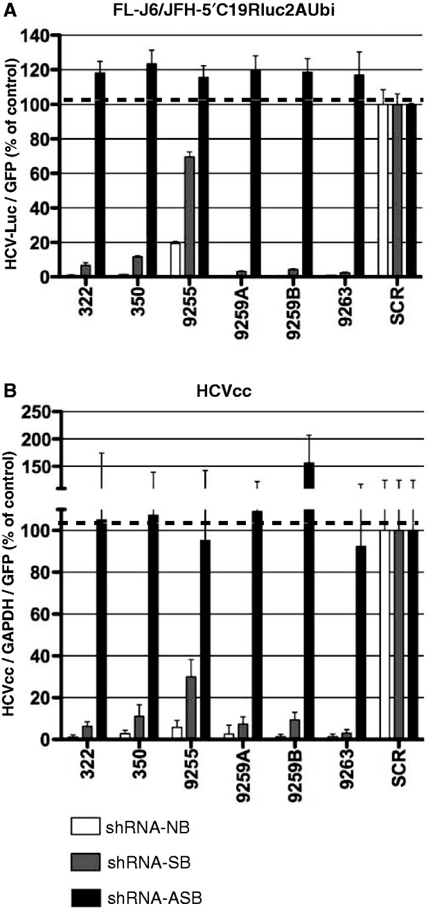
shRNA without bulges (NB-no bulge, white bars) and asymmetric shRNA with bulges in sense strand (SB-sense bulge, gray bars) or antisense strand (ASB-antisense bulge, black bars) were tested against replicating FL-J6/JFH-Luc HCV (A) and HCVcc infection (B) in Huh7.5 cells. SCR shRNA was used as the control and assigned a value of 100. In (A), the luciferase signal was normalized to GFP expressed from the AAV-shRNA vectors to standardize for the transduction efficiency. In (B), quantitative PCR was used to detect HCV levels, and values were normalized to GAPDH quantitative PCR levels as well as AAV-shRNA vector encoded eGFP. Error bars represent SD from n = 3–6.