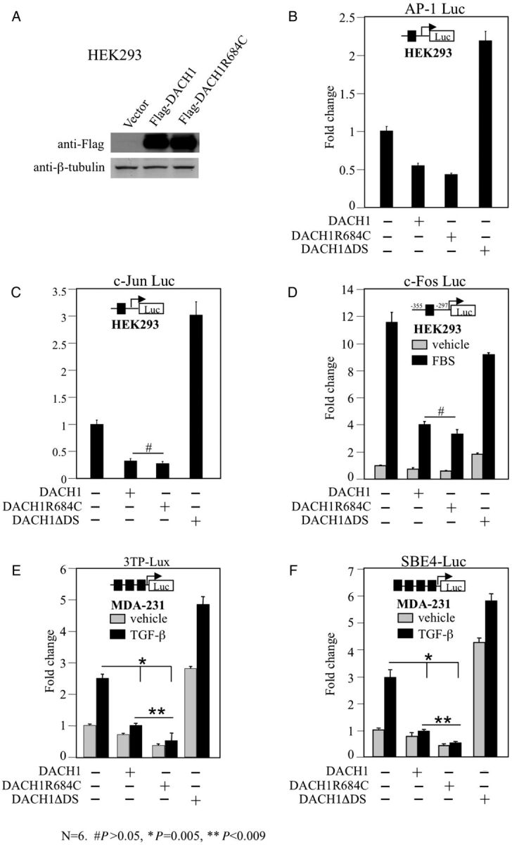
Fig. 3.

Repression of TGF-β signalling by DACH1-p.R684C. Western blot analysis of HEK293T cells transiently transfected with Flag-tagged DACH1 and DACH1-p.R684C expression plasmids demonstrating equal protein abundance of wild-type and mutant DACH1 (A). Repression levels of AP-1 (B), c-Jun (C) and c-fos promoter (D, in response to serum stimulation) by wild-type DACH1 and DACH1-p.R684C in HEK293T cells. DACH1 protein with deleted DS domain (DACH1ΔDS) served as negative control. Strong repressor activity of DACH1-p. R684C in MDA-231 cells after co-transfection with 3TP-Lux (E) and SBE-4 Luc (F) reporter and stimulation with or without TGF-β for 12 h. DACH1 protein with deleted DS domain (DACH1ΔDS) served as negative control. Data are shown as mean ± SE for n = 6 separate transfections.
