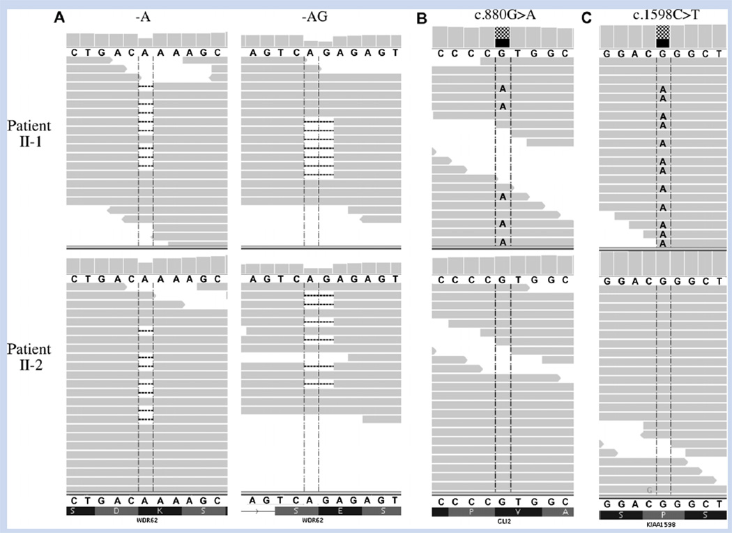
FIG. 2.
A: Sequence alignment visualization showing heterozygous WDR62 mutations in both patient II-1 (upper panel) and patient II-2 (lower panel). Individual sequencing reads are represented by the horizontal grey bars. Both c.2083delA and c.2472_2473delAG deletions are present in approximately half of each patient’s reads, representing a heterozygous state for each mutation. Alignment also shows heterozygous missense variants in (B) GLI2 and (C) KIAA1598 in patient II-1 but not in patient II-2. [Color figure can be seen in the online version of this article, available at http://onlinelibrary.wiley.com/journal/10.1002/(ISSN)1552–4833]

